
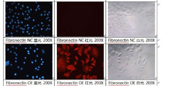
利用荧光标记抗体与目的抗原的特异性结合,通过荧光显微镜对抗原处的荧光进行拍照 ,可知细胞中目的抗原的定位和表达情况。
根据抗原抗体反应的原理,先将已知的抗原或抗体标记上荧光基团,利用这种荧光抗体(或抗原)作为探针检查细胞内的相应抗原(或抗体)。借助荧光显微镜观察荧光,从而确定细胞内抗原/抗体的性质,并对其进行定位。比较不同条件处理的实验组之间,细胞内蛋白的表达差异和细胞定位。
目的细胞按实验设计组别进行药物处理后,收集细胞悬液,离心,用缓冲液洗涤,加细胞染色缓冲液重悬后,用PE标记的CD11b抗体进行染色,染色结束后,用细胞染色缓冲液洗涤、重悬,进行流式仪检测 (Millipore,Guava easyCyte HT),分析各实验组别中CD11b 阳性细胞百分比差异。
1.COONS AH, KAPLAN MH. Localization of antigen in tissue cells; improvements in a method for the detection of antigen by means of fluorescent antibody. J Exp Med. 1950 Jan 1;91(1):1-13.
2.Gobert GN. et al. Immunolocalization of NuMA and phosphorylated proteins during the cell cycle in human breast and prostate cancer cells as analyzed by immunofluorescence and postembedding immunoelectron microscopy. HistochemCell Biol. 2001 May;115(5):381-95.
3.Petrek M. Identification of lymphocyte subpopulations by indirect immunofluorescence--a micromethod. Acta Univ Palacki Olomuc Fac Med. 1991;129:51-6.4/ 4
4.Stănculescu R. et al. Immunofluorescence expression of Ki-67, p53 and cyclin inhibitors (p16ink4a, p21 and p27) in low-grade cervical lesions versus high-grade cervical lesions. Research study on cell cultures. Rom J Morphol Embryol. 2013;54(3Suppl):725-34.
5.Pollex T. et al. Live-cell imaging combined with immunofluorescence, RNA, or DNA FISH to study the nuclear dynamics and expression of the X-inactivation center.Methods Mol Biol. 2013;1042:13-31.


