The development of chick embryos has been studied since Aristotle. It is one of the most intensely studied organisms. One reason for this is that there are great similarities between avian and mammalian embryology. Another advantage is that the embryo is relatively accessible and can be operated on or treated with teratogens (substances that perturb development) while still in ovo(in the egg). Finally, fertilized chicken eggs are available year round and can be "held" at 10-14ºC for several weeks before being set, producing embryos upon demand.
Embedded embryos
1. Examine plastic embedded specimens of "13-hour" or "16-hour", "24-hour", "33-hour","48-hour" and "72-hour" chick embryos. These terms refer to classic studies of chick development. In reality, chick embryos will develop at different rates for a number of reasons, including incubation temperature. In modern times, chicks always take longer to reach these stages. Two more accurate ways of describing the relative developmental age of a chick embryo are (1) the number of somites and (2) comparison to a staging series, such as the one devised by Hamburger and Hamilton (H&H; hamburger.pdf). The somites are the blocks of tissue on either side of the neural tube. Try to determine the number of somites and the H&H stage number of your embryos.
2. Try to identify the structures indicated in the diagrams. Pay particular attention to the developing circulatory system. Try to determine which germ layer forms each structure. Highlights include:
13- to 16-hour.Identify the area opaca and area pellucida. Locate the primitive streak. Hensen's node is located at the anterior end of the streak.
24-hour.Locate the neural folds and Hensen's node. Identify the anterior end of the embryo.
33-hour.The embryo is lying along the center of the blastodisc, dorsal side up. The heart is to the right, along the side of the hindbrain. It is a simple, looped tube;connected to the vitelline vein and the ventral aorta. What is the function of the vitelline vein? Identify the forebrain, midbrain and hindbrain. The optic vesicles are outpockets on either side of the forebrain. The neural plate runs along the dorsal midline and is not yet closed. You may also be able to see the notochord running along the midline.
48-hour.The head has bent forward and the body has begun to twist. The optic cup(eye) and the otic vesicle (ear) have formed. The neural tube has closed, except in the region of the hindbrain. A series of aortic arches run between the pouches
in the pharynx and converge into the dorsal aorta. The heart has begun to differentiate into an atrium and a ventricle.
72-hour.The flexure of the embryo is now pronounced and the embryo is lying on
its left side. The brain has further subdivided and the olfactory pit (nose) is
present over the tip of the forebrain. The lens of the eye should be visible.
Lateral swellings representing the limb buds and a curved tail are also present.
Living embryos
1. Isolate a living 3- to 6-day chick embryo. Obtain an egg and clean scissors, forceps,
a plastic spoon. First prepare a small petri dish withHoward Ringer's solution.
Gently rotate egg, clean with 70% ethanol and set down for a few minutes to
allow the embryo to float to the top. Open up the blunt end of the shell and
carefully peel back the shell membranes. Observe the embryo under the
dissecting scope.
2. Measure the heart rate and observe any other movement. Grab
the outer ring of the blastodisc near embryo with fine forceps in your non-cutting
hand. Try not to pinch any blood vessels. Quickly cut around embryo.
3. Keeping hold of the embryo with the forceps, pick up a plastic spoon with your
cutting hand and slide it under the embryo. Continue to hold onto embyo, lift
with the spoon and transfer to the petri dish filled with Howard's and examine
with the dissecting microscope. have your lab partner stand by with scissors in
case the embryo wasn't completely detached. If the dish is cloudy with yolk,
transfer embryo to a fresh dish of Howard's.
Note: an alternate method of embryo isolation is to break the egg open into a finger bowl of Howard's. This is similar to cracking an egg for culinary purposes, but the yolks of warm eggs are more fragile than those of refrigerated ones. First, place the egg in a horizontal position to allow the embryo to float to one side. Next, keeping the same orientation, crack the egg gently against the side of a finger bowl filled with Howard's. Submerge the egg and gently pull the ends appart to crack the egg open.

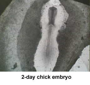
4. Isolate a 1- or 2-day embryo. Clean your dissecting equipment and prepare a fresh
dish of Howard's. Clean an egg and allow the embryo to float to the top.
Open the blunt end of the egg and remove the shell membranes. the embryo
may not be visible to the naked eye. The blastodisc is located above a small ring
of white yolk. Remove albumin with wide-mouth pipet and shell with forceps.
5. Drop a filter paper disc around your embryo. Hold on to the filter
paper with fine forceps and cut around the ring with your sharp scissors.
Transfer the embryo to a small petri dish with Howard's Ringer's solution.
6. Examine both embryos. Pay particular attention to the heart and circulation, and
to the developing neural tube. Compare the heart rate between the 2 embryos.
Which side was towards the yolk? The reddish spots on the 2-day blastodisc are
the blood islands, the sites of hematopoesis. The embryo is covered with a clear
protein layer known as the vitelline membrane. This may start to peel away
from the embryo.
7. For each embryo,determine the H&H stage. How do these embryos compare to
the stained specimens?
8. Clean your instruments well with warm water, distilled water and 70% ethanol.
Dry before returning to case. Discard the shells and the yolk/albumin remains.
|
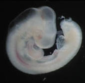
3-day chick embryo |


 首页
首页