引言:上海吉凯基因化学技术有限公司成立于2002年,拥有国内最大的慢病毒文库,同时也是国内最大的疾病关键基因科研服务供应商,在疾病研究领域的实验积累已逾14年!在这里,我们为大家特别整理了10多个研究领域的经验所得,将最实用的精华分享给大家!上一期帮大家总结了肺癌研究中慢病毒工具的应用,上期回顾看这里本期我们来看看胃癌又有哪些需要注意的地方呢。
胃癌具有高度异质性,通过常规病理可以将胃癌进行一定的区分,但目前广泛应用的组织病理学分型已逐渐难以适应临床个体化诊疗的需求,对于胃癌的研究已进入分子水平。俗话说“良好的开端是成功的一半” 。今天就给大家聊一聊胃癌研究中,体内外模型的选择,以及病毒工具的应用。

一、我需要哪种细胞模型?是否易于操作?
大量的组学研究已经证实了胃癌是一种非常复杂的疾病,如果您想要研究某种特殊分群的胃癌,有两个问题一定要注意:1.所用的细胞、动物模型是否符合疾病的特征;2.如何在模型中根据实验需要,对目的基因进行有效的操作。举个栗子,如果想要研究胃癌中某个基因对p53通路的影响,需要选择什么样的细胞呢?
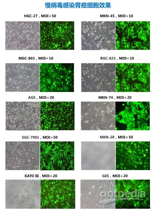
来来来,这里有胃癌各种类型的常用细胞系,每株细胞都配有吉凯独家的预实验数据哦,快来吉凯的表里找找吧~

*更多更全的预实验信息,可咨询当地业务员
二、细胞实验效果不错,那动物实验怎么做?
胃癌研究中动物实验主要分为三种类型:I自发胃癌模型;II诱导胃癌模型;III 移植胃癌模型。第I种主要是通过构建转基因动物的方式实现,第II种则利用化学诱导剂或幽门螺旋杆菌 (HP)感染诱导胃癌发生,第III种将人类肿瘤组织或细胞直接接种于裸鼠进行研究。因裸鼠培育条件低,无毛,易于动态观察肿瘤的生长状态,且通过接种方式、部位的选择可达到较满意的转移效果,因此目前多选择裸鼠进行人胃癌细胞异体移值。

对于移植瘤模型中的基因操作,较为常见的是慢病毒感染细胞后接种。若是选择病毒直接注射动物/瘤体,所用的病毒需经过特殊纯化处理,确保滴度高,毒性小,不易引起炎症反应。

三、体内外模型选好了,那么我的实验需要使用哪种慢病毒载体对基因进行操作呢?
启动子、荧光标记、抗性标签是我们选择载体需要考虑的三大要素。实验不同,这些元件也需要相应变化。祭出神图一张,定制你的专属载体,就是这么简单!
参考文献
【1】Yokozaki, Hiroshi. “Molecular characteristics of eight gastric cancer cell lines established in Japan.” Pathology international 50.10 (2000): 767-777.
【2】Li, Wenjuan, et al. “CIP2A is overexpressed in gastric cancer and its depletion leads to impaired clonogenicity, senescence, or differentiation of tumor cells." Clinical Cancer Research 14.12 (2008): 3722-3728.
【3】Wang, Jia-bin, et al. “CDK5RAP3 acts as a tumor suppressor in gastric cancer through inhibition of β-catenin signaling.” Cancer Letters (2016).
【4】Sun, Ruochuan, et al. “Down regulation of Thrombospondin2 predicts poor prognosis in patients with gastric cancer.” Molecular cancer 13.1 (2014): 1.
【5】Zhang, Qi, et al. "Knockdown of S100P by lentiviral-mediated RNAi promotes apoptosis and suppresses the colony-formation ability of gastric cancer cells." Oncology reports 31.5 (2014): 2344-2350.
【6】Zhang, Qing, et al. "By recruiting HDAC1, MORC2 suppresses p21Waf1/Cip1 in gastric cancer." Oncotarget 6.18 (2015): 16461.
【7】Zhou, Jinfeng, et al. “Chaperone-mediated autophagy regulates proliferation by targeting RND3 in gastric cancer.” Autophagy 12.3 (2016): 515-528.
【8】Li, T., et al. "MicroRNA-296-5p increases proliferation in gastric cancer through repression of Caudal-related homeobox 1." Oncogene 33.6 (2014): 783-793.
【9】Asciutti, Stefania, et al. “Diverse mechanisms of Wnt activation and effects of pathway inhibition on proliferation of human gastric carcinoma cells.” Oncogene 30.8 (2011): 956-966.
【10】Zhou, W., et al. “The AKT1/NF-kappaB/Notch1/PTEN axis has an important role in chemoresistance of gastric cancer cells.” Cell death & disease 4.10 (2013): e847.
【11】She, Jun-Jun, et al. “Side population cells isolated from KATO Ⅲ human gastric cancer cell line have cancer stem cell-like characteristics.” World J Gastroenterol 18.33 (2012): 4610-4617.
【12】Zhang, Y., et al. “SIRT1 Is Reduced in Gastric Adenocarcinoma and Acts as a Potential Tumor Suppressor in Gastric Cancer.” Gastrointestinal Tumors 2.3 (2015): 109-123.
【13】Su, Guo‑Qiang, et al. “Research of shRNAmir inhibitory effects towards focal adhesion kinase expression in the treatment of gastric cancer.” Oncology letters 9.2 (2015): 595-603.
【14】Hu, Hao, et al. “Real-time bioluminescence and tomographic imaging of gastric cancer in a novel orthotopic mouse model.” Oncology reports 27.6 (2012): 1937.
【15】Chen, L., et al. “miR-1207-5p and miR-1266 suppress gastric cancer growth and invasion by targeting telomerase reverse transcriptase.” Cell death & disease 5.1 (2014): e1034.
 首页
首页