Isolation of mice glomeruli
1. Mice were anesthetized by an
intraperitoneal injection of Avertin (2,2,2-tribromoethyl and tertiary
amyl alcohol; 17 μl/g mice) and perfused with 8 × 10⁷ Dynabeads diluted in 40 ml of phosphate-buffered saline through the heart.
2. The kidneys were removed, minced into 1-mm3
pieces, and digested in collagenase (1 mg/ml collagenase A, 100 U/ml
deoxyribonuclease I in HBSS) at 37°C for 15 minutes (for newborn mice)
or 30 minutes (for adult mice) with gentle agitation.
3. The
collagenase-digested tissue was gently pressed through a 100-μm cell
strainer using a flattened pestle and the cell strainer was then washed
with 5 ml of HBSS.
4. The filtered cells were passed through a new cell strainer without pressing and the cell strainer washed with 5 ml of HBSS.
5. The cell suspension was then centrifuged at 200 × g for 5 minutes.
6. The supernatant was discarded and the cell pellet was resuspended in 2 ml of HBSS.
7. Finally,
glomeruli containing Dynabeads were gathered by a magnetic particle
concentrator and washed for at least three times with HBSS.
8. During the procedure, kidney tissues were kept at 4°C except for the collagenase digestion at 37°C.
Morphological Studies
1. Dynabead-perfused kidneys were
snap-frozen for cryostat sectioning. Sections were stained with
hematoxylin and eosin (H&E) and were examined by light microscopy.
2. Isolated
glomeruli were examined by both light and electron microscopy (EM).
Specimens for EM were fixed with 2% paraformaldehyde and 2.5%
glutaraldehyde.
3. Glomeruli intended for transmission EM were
subjected to ferrocyanide-reduced OsO4, dehydration, and plastic
infiltration before ultrathin sectioning.
4. For scanning EM glomeruli were osmicated according to the OTOTO protocol and dried using hexamethyldisilazane evaporation. 
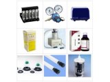
Flow chart of the isolation technique of mice glomeruli.
Reference
1. Friedman PL, Ellisman MH: Enhanced visualization of
peripheral nerve and sensory receptors in the scanning electron
microscope using cryofracture and osmium-thiocarbohydrazide-osmium
impregnation. J Neurocytol 1981, 10:111-131.
2. Braet F, Dez Anger
R, Wisse E: Drying cells for SEM, AFM and TEM by hexamethyldisilazane: a
study on hepatic endothelial cells. J Microsc 1997, 186:84-87.


 首页
首页


