Sometimes it's not enough to know how much drug has reached a particular tissue.
Sometimes you need to know where in that tissue the drug has gone.
To answer that and other questions, pharmaceutical companies are turning to imaging mass spectrometry. It allows pharma scientists to collect information about drug distribution much earlier in the discovery process than previously possible.
The resulting pictures are helping drug developers prevent toxicity and off-target effects.

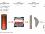
figure: MS imaging helps visualize earlystage fibrosis in a rat liver (top) by showing the location of hepatocytes (blue), fibrotic bands (green), and the portal vein (red). In the center image, the MS data are overlaid on the conventional histology image (bottom).


 首页
首页