Drosophila Schneider S2 cells
Schneiders Medium (GIBCO/Invitrogen), 10% fetal calf serum, Antibiotics (Sigma A5955)
Depression slides (Karl Hecht GmbH)
Coverslips No. 1.5 (thickness on average 170 µm) are optimal for the Olympus™ microscopes and also recommended by other microscope manufacturers.
Culture slides/dishes (ibidi®, Lab-Tek™ Chambered Coverglass/Nunc™, Wellco Wells®)
Optional: 0.1% Poly-L-Lysine Stock (Sigma, P8920)
Optional: Concanavalin-A (Sigma, C2010-25MG)
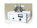
Deltavision Spectris® and Deltavision RT® system using an inverted Olympus™ IX71 microscope.
Filter sets from Chroma™ or Semrock™ (Sedat filter set).
Temperature controlled environmental chamber from Solent Scientific™, PeCon™, Zeiss™ or Olympus™.
Image analysis software used (softWoRx® Explorer, Applied Precision™, Bitplane Imaris®, Metamorph®, Microsoft Excel™).
Back to top
Cells keep dividing under these conditions for at least a day. For longer periods of time, cells should be grown in culture dishes with coverslip bottoms (see Protocol B for long periods of time).
Schneider S2 cells are semi-adherent, so specific treatment of the coverslips is not required. If cells should be immobilized further, acid-washed coverslips can be coated using 0.01% Poly-L-Lysine Solution (see Protocol B). If flattened cells are desired, acid-washed coverslips should be coated with Concanavalin-A. Acid washed 22x22mm coverslips that are treated with 100µl 0.5mg/ml concanavalin A (Sigma) in water and allowed to air dry (Rogers et al., 2002). However many cells fail to complete cytokinesis and become bi-nucleated under these conditions.
In all these experiments stable conditions around the microscope are mandatory as slight deviations in temperature can change the focus dramatically. This can be achieved separating the microscope from strong airflow (e.g. in climatized rooms through curtains - also useful to create a dark partition of the room) and the use of an environmental chamber that encloses most of the microscope. A constant temperature in a temperature-controlled chamber can only be guaranteed if the microscope room is at least 5°C lower than the chamber temperature. Alternatively, but less precise is the use of a heated stage with temperature control. Unless the environmental chamber can be both cooled and heated, the above-described setup almost always requires an air-cooling system in the room.
Culture wells mounted on slides have the advantage, that slides can be positioned in a reproducible manner on the microscope stage. This allows taking the slide on and off and identifying previously observed cells by coordinates.
The use of the Olympus™ Planapo 60x objective is preferable over the 100x objective, because it is more light sensitive yet provides a decent optical resolution to visualize substructures in the cell (chromosomes, nuclear bodies, microtubules).
Back to top
Reviewed by: Helder Maiato, Institute for Molecular and Cell Biology, University of Porto, Portugal.
The use of the hanging drop method to leave oxygen available may just be important for long-term recordings. An alternative for short-term recordings (up to 6 hours) would be to use closed chambers (like Rose chambers) full with medium.
An alternative to Poly-L-Lysine, which just helps to stick S2 cells to the glass, and to Concanavalin-A, which flattens cells by using membrane receptors and may compromise cytokinesis, is to use S2R+ cells. This clone was originally isolated by Imogene Schneider (Schneider, 1972) and grows adherently on glass without any coating and as a loose monolayer, predominantly epithelial-like in appearance. As opposed to regular S2 cells, this clone expresses both Wingless receptors Dfrizzled-1 and Dfrizzled-2, hence the new nomenclature of "S2 receptor plus" (S2R+; Yanagawa et al., 1998). Another alternative to image flat S2 cells for short periods (2-3 hours) is to use an agar overlay (Maiato et al., 2004). For some purposes, this method results better than all the others, since it works based on the surface tension of the fluids and a pressure gradient that allows the visualization of cells that are flat enough for microscopy, without compromising cell viability.


 首页
首页


