Moreover, when the method was applied to the analysis of the cellular composition of immature testes, the results were also in agreement with previous reports. As an example, the relative percentages obtained by our method for testicular subpopulations differing in DNA content (C, 2C, and 4C) for 21-day-old rats, were 0%, 65%, and 24%, respectively (Fig. 2 d). The corresponding previously reported values were 0%, 77%, and 19% for 20-day-old rats, and 0%, 48%, and 47% for 22-day-old rats (9). Therefore, we have obtained intermediate relative percentages of C, 2C, and 4C cells for an intermediate developmental stage, which would again indicate that our method does not selectively damage any specific cell type.
Thus, the viability and cell type proportions in the cellular suspensions obtained with the mechanical method described here do not significantly differ from the previously reported results using more time-consuming and laborious approaches. Besides, the reproducibility of the method was very high, specially if individual variations are taken into account (Table 3 ).
Table 3 Reproducibility analysis of the method described here
Rattus norvegicus specimen | DNA content | ||
|---|---|---|---|
C | 2C | 4C | |
1 | 72.0 | 12.0 | 16.0 |
2 | 76.0 | 11.0 | 13.0 |
3 | 75.0 | 13.0 | 12.0 |
4 | 76.0 | 11.0 | 13.0 |
5 | 72.0 | 12.0 | 16.0 |
6 | 71.5 | 15.0 | 13.5 |
7 | 73.0 | 13.0 | 14.0 |
Arithmetic mean | 73.6 | 12.4 | 14.0 |
Standard deviation | 2.0 | 1.4 | 1.5 |
Relative percentages of C, 2C, and 4C cell populations from seven independent experiments performed with adult rats (one individual per experiment) are shown as an example
Summarizing, we have developed a very simple, fast, and reproducible cell suspension-preparation method for flow cytometry analysis of rodent testicular cell populations. Although not tested here, this method could be also used in combination with other cell purification techniques such as elutriation or staput. Additionally, since this method involves very little manipulation and avoids enzymes and detergents, it could become an ideal choice for delicate downstream applications such as gene expression studies. In this regard, we are now using sorted spermatogenesis cell populations (see supplemental Fig. 1, accessible at http://www.iibce.edu.uy/PAPERLINKS/RRCasuriaga-supplemfig1.jpg) for reverse transcriptase-polymerase chain reaction amplification of stage-specific transcripts (to be published elsewhere). Although we have not tested the preservation and/or specificity of any spermatogenic protein yet, our method could also be advantageous for protein studies considering the absence of trypsin in the procedure.
In conclusion, time savings, little manipulation involved, and the quality of the resulting suspension make this method an interesting alternative to the more tedious and time-consuming cell preparation techniques currently in use.
Acknowledgements This work was supported by DYNACYT (Fondo Clemente Estable, Project N° 10171). The authors are indebted to Dr. Mariela Bollati for her generous collaboration concerning the MoFlo cell sorter and Federico Santiñaque for cytometric data handling. We also wish to thank Merial-Montevideo for gently providing all the guinea pig specimens used in this work as well as Marcelo Fernández for animal technical assistance.
Appendix
Protocols
Preparation of cell suspensions
Materials:
| – | Glass Petri dishes |
| – | Scissors and forceps |
| – | Medimachine (BD) |
| – | Medicon units (BD) |
| – | 50 μm nylon membrane or 50 μm Filcon units (BD) |
| – | 25 μm nylon membrane or 25 μm Filcon units (BD) |
| – | 5 mL syringes |
| – | Neubauer chamber |
| – | Separation medium: Dulbecco’s Minimal Essential Medium (D-MEM ) supplemented with 10% fetal calf serum |
| – | NDA (2-naphthol-6,8-disulfonic acid, dipotassium salt) |
Procedure:
| 1. | Place the dissected testis in a 96-mm glass Petri dish on ice, containing 10 mL of ice-cold separation medium. |
| 2. | Remove the tunica albuginea and cut the decapsulated testis into 2–3 mm3 pieces. |
| 3. | Place four to five of these pieces in a disposable disaggregator Medicon™ (BD) along with 1 mL of cold separation medium and process in the Medimachine system for 50 s. |
| 4. | Recover the resulting cell suspension using a 5-mL syringe without needle. |
| 5. | Filter through a 50-μm nylon mesh [or Filcon™ unit (BD) containing a similar mesh], previously soaked with 0.5 mL of separation medium. |
| 6. | Repeat step 5 but using a 25-μm nylon mesh (or equivalent BD Filcon™ unit). |
| 7. | Take an aliquot of the cell suspension to count in a Neubauer chamber and adjust cellular concentration to 1–2 × 10⁷ cells/mL. |
| 8. | Add NDA to a final concentration of 0.2% to avoid cell clumping. |
Cell viability evaluation
Materials:
| – | LIVE/DEAD viability kit for animal cells (Molecular Probes, Eugene, OR, USA) |
| – | Trypan blue |
Procedure:
Check cell viability of the testicular cell suspensions with the LIVE/DEAD viability kit for animal cells (Molecular Probes, Eugene, OR, USA) following manufacturer’s instructions. Alternatively, trypan blue dye exclusion test can be performed as detailed below:
| 1. | Take 0.1 ml of the concentrated cell suspension and dilute it to an approximate concentration of 1-2 × 10⁵ cells/mL. |
| 2. | Add 0.1 ml of 0.4% trypan blue stain to 0.5 mL of the diluted suspension. Mix thoroughly. |
| 3. | Allow to stand 5 min at room temperature. |
| 4. | Fill a hemocytometer as for cell counting. |
| 5. | Under a microscope, count nonviable (stained) and viable (unstained) cells. |
Flow cytometry analysis
Materials and equipment:
| – | Hoechst 33342 (Sigma-Aldrich, St. Louis, MO, USA), stock 5 mg/mL |
| – | MoFlo Cytometer (DakoCytomation) equipped with a UV excitation wavelength laser (Innova 90C-6) |
Procedure:
| 1. | Prior to flow sorting, add Hoechst 33342 to the cell suspension to a final concentration of 5 μg/mL. Incubate for 10 min at 37°C in the dark. |
| 2. | Perform cell analysis by means of a MoFlo Cytometer (DakoCytomation) equipped with a UV excitation wavelength laser (Innova 90C-6) operating at 25 mW. Prior to cell analysis, check instrument linearity and doublet discrimination performance with DNA QC Particles (Becton Dickinson) stained with Hoechst 33342. |
| 3. | Use Summit v4.3 software (or a similar one) to analyze the following parameters: forward scatter (FSC-H); side scatter (SSC-H); pulse-area or total emitted fluorescence (FL2-A); and pulse-high or intensity of fluorescence emission (FL2-H). |
Electronic Supplementary Material
Below is the link to the electronic supplementary material.
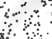
Supplemental Figure 1 Partial view of sorted 4C guinea pig cells. The cell suspension was stained with Hoechst 33342 (10 min) and analyzed with a MoFlo cytometer (DakoCytomation) equipped with a UV excitation wavelength laser (Innova 90C-6) operating at 25 mW and a 70-μm nozzle. Sorted cells were recovered onto 12 × 75 mm polystyrene tubes, centrifuged at 450 g (5 min) and 0.5 ml of paraformaldehyde (1%) were added to the cell pellet. Aliquots of fixed cells were dropped onto clean microscope slides and nuclei stained with Giemsa (3%, 5 min). Note the similarity of nuclei size and morphology (GIF 1573 kb)


 首页
首页


