Objective:The overall objective of this laboratory experiment is to examine specification in the axolotl through transplantation of the gill organ field.
Introduction:The axolotl (Ambystoma mexicanum) is a large salamander which is native to Lake Xochimilco, Mexico. The axolotl is a member of a larger group of salamanders known as mole salamanders. Other members of this group include tiger salamanders and spotted salamanders. The wild type axolotl is distinguished by its dark color which may contain greenish mottling and silvery patches. The albino type axolotl is white (without pigment). The average axolotl can reach a length of approximately 12 inches from nose to tail and can weigh up to 300 grams. Axolotls are neotenic, which means they will possess their feathery external gills and tail fin their entire lives while they maintain an aquatic lifestyle
Background:The goal of this experiment is to determine if the gill organ field is specified at the particular time that it is transplanted. According to Gilbert (2000), a cell or a tissue is said to be specified when it is capable of differentiating as it would have when placed in a neutral environment such as a petri dish or test tube or after transplantation to an ectopic site. An organ field is a large region of the embryo in which cells are specified generally (leg, gill) but are not specified for a specific organ such as a toe or finger. Organ fields are developed in the axolotl through induction. An interaction at close range between two or more cells or tissues that have different properties is called induction (Gilbert, 2000). There are two components necessary for inductive interaction. The first component is the inducer, which is the tissue or cells that produce a signal(s) that will change the cellular behavior or activity of the other cells or tissue. The second component is the responder, which is the tissue or group of cells that are being induced (Gilbert, 2000). Coordination in the construction of organ fields is achieved as one group of cells changes the behavior of an adjacent set of cells, which will cause the cells to change shape, mitotic rate, or fate (Gilbert, 2000). Once the organ field is specified, inductive effects will help further develop the field into the final organ. By transplanting the gill organ field from a wild type axolotl into an albino axolotl and vice versa, we plan on examining if the gill develops as it would have in its original environment or if it is influenced by the surrounding tissues. We expect the gill to develop as it would have in its original environment.
Procedure:
1. Obtain axolotl embryos. For this experiment, we will be using wild type and albino axolotl embryos at stage 21 in their staging series.
2. Prepare the axolotl embryos as follows:
a. manually dejelly embryo of appropriate stage and keep inHEPES-buffered Modified Steinberg's solution (HBSt) containing antibiotics until use. For microsurgery, also remove membrane around embryo.
b. place embryos into operating dish containing 1 x HBSt and antibiotics
c. use clean technique; dip tools, pipettes, and glass bridges in 70% EtOH and then into sterile HBSt before using
3. Start microsurgery on your donor embryo. Locate the gill organ field which is in the first anterior third of the embryo (toward head) and right above the small crest or indention closer to the ventral side (Rugh, 1962) . Isolate the gill organ field (or the region that will become the gill organ field) by cutting around it and detaching that specific piece of tissue from the embryo.
4. Start transplantation on your recipient embryo. Cut a small slit in a region close to the gill organ field on the recipient. Gently slip donor gill organ field into slit and be sure to secure the graft in place. It is critical to try to get the graft as far under the cut as possible to ensure that the recipient will begin to incorporate the new tissue into its development process.
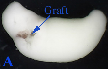
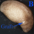
Figure 1:
A. A 21 stage albino axolotl embryo with a transplanted wild type axolotl graft (supposedly the gill field). The picture was taken immediately after microsurgery.
B. A 21 stage wild type axolotl embryo with a transplanted albino axolotl graft. The picture was taken immediately after microsurgery
5. Once the surgery is complete, be sure to take pictures of a normal donor and recipient and then the newly transplanted graft of the recipient. Pictures should be taken at various stages to observe development (to be determined at discretion of researcher).
6. The embryos should be left in HBSt at room temperature to continue to development. Observe both donors and recipients as time continues to examine the effect of the transplanted graft.
7. Make OBSERVATIONS as development continues...this is the key to obtaining results!
8. After the first day, it is important to replace the operating solution with 10% modified Steinberg's solution. The graft will develop better in a lower concentration of certain chemicals. To do this, simply pipette off the operating solution and pour in the 10% modified Steinberg's solution.
Results/Discussion:Grafts were successfully transplanted into donor recipients. A wild type axolotl that had received an albino transplant survived the longest and displayed the ability to retain the transplanted graft and continue development. The albino recipients of wild type grafts were not as successful because the graft blended into the albino's natural color and could not be easily distinguished. Microsurgery was complicated and the regions that were transplanted either were not the gill organ field or were not directly transplanted into the gill organ field. However, it is important to note that those grafts that were transplanted did successfully stay in the recipients and the recipients continued to develop with the graft.

Figure 2: A picture of a wild type axolotl embryo 2 days after the albino axolotl graft had been transplanted into it. The graft is located below the gill field but still has been successfully transplanted and has become a part of the developing embryo.
We expected that the transplanted graft would have developed as if it were a part its original environment. This implies that the gill would have been specified prior to transplantation. Upon transplantation, inductive effects would allow the transplanted graft to eventually form the gill. There are two reasons why we might not have examined any of our expected results. The first is the possibility that the gill organ field had not been specified prior to transplantation, which did not allow it to develop properly when it was transplanted into the recipients. The second reason is due to technology. This was a very complicated experiment that required skilled microsurgery on embryos. Since this was a first attempt at microsurgery, our technique and technology might have been insufficient which caused poor results to be obtained.


 首页
首页


