The Effects of NiCl2on Spicule Formation
Jessica Ann Billet, Franklin and Marshall, Class of 2000
Background and Objective
Sea urchins exhibit radial holoblastic cleavage, eventually forming a blastula. Shortly after the blastula hatches from the fertilization membrance, the embryo begins gastrulation. Gastrulation begins when the vegetal side of the blastula begins to thicken and flatten. This flat sheet of cells is called thevegetal plate. In the center of the vegetal plate a small group of cells begins to change. These cells extend and contract long, thin filopodia. These cells then break off from the epithelium and migrate into the blastocoel. These migrating cells are known as the primary mesenchyme cells (PMCS). Eventually the migrating cells localize within the ventrolateral region of the blastocoel. It is in this area that the PMCs fuse together to form syncytial cables. Syncytial cables will eventually form the axis of the calcium carbonate spiculesof the larval skeleton.
The purpose of this lab is to explore the development of spicules in a sea urchin's larval skeleton. NiCl2interferes with spicule and skeletal formation, by introducing half of the embryos into a solution of NiCl2, we hope to observe the effects of blocking skeleton formation. We will stain all of the embryos with an Ig8 immunoflourescent antibody. Ig8 will stain or 'tag' the primary mesenchyme cells of the developing embryo.
This lab will take two weeks to accomplish. In the first week, we will collect and fertilize sea urchin gametes. One half of the embryos produced will then be subjected to a NiCl2solution until Friday morning. On Saturday, the embryos will be fixed in preparation for staining in week two. During lab next week we will stain the labs with Ig8 antibody, an immunofluorescent stain. We will use this stain to identify PCMs and developing spicules.
** We will be usingHytechinus variegatusfrom the Florida coast.The gametes and embryos can be left at room temperature in order to develop**
Experimental Protocol
A. Fertilization of Urchins
See standard protocol.
B. NiCl2Treatment
1)After fertilization has occured, transfer half of the embryos into a solution of 10mM NiCl2. Transfer the remaining half, the control, into a solution of ASW. 2)The two groups should be left at least 24 hours to develop. Track the development through the mesenchyme blastula stage.
3)The groups should be prepared for stain when the control group (ASW) reaches gastrulation and has formed the pluteus larva.
C. Staining
See standard protocol.
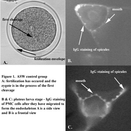
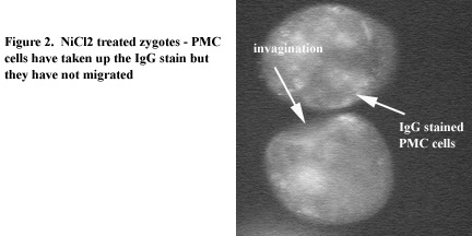
Results and Discussion The two samples behaved quite differently. The Ig8 staining revealed that PMCs of the control group had in fact migrated after gastrulation to form the endoskeleton, or spicules, of the urchin. In the experimental group, none of the embryos had undergone complete gastrulation. The Ig8 staining showed that the PMC cells had failed to migrate out, the embryo itself had failed to develop. |
References:
- Influence of NiCl2on the Skeleton Formation in Sea Urchin:
Armstrong et al. (1993) Development 119:833-840;
Hardin et al. (1992) Development 116:671-685.
- Fertilization And Early Development Of The Sea Urchin.
Cebra-Thomas, Judy. Spring 1998.
- Basics of Sea Urchin Development.
Cebra-Thomas, Judy. Spring 1999.
Gilbert SF. editor. 1997. Developmental biology. 5th ed.: Sinauer Associates, Inc., Sunderland, Mass, 918 p.


 首页
首页