摘要:基于抗体的蛋白质检测方法,主要有western blot、ELISA、点杂交以及免疫组化等,这些方法被广泛地应用于科研和诊断领域。在蛋白质的免疫检测过程中,样品蛋白首先结合与特异性一抗上,然后再用携带标签(诸如:荧光染料,放射性元素,酶等)的二抗进行检测。然而,为了避免种间内的交叉反应,必须谨慎地选择二抗。同时,有限的、可利用的一抗和二抗抗体对,进一步限制了对蛋白质的免疫检测。而本文主要介绍一种不需要二抗的、基于DNA的蛋白质检测系统,该系统主要是应用一种既和DNA修饰的报告分子结合,又与IgG抗体结合的通用型适配子,替代了标记二抗,可以对单一实验进行多重检测,并实现了功能的模块化。文中以DNA修饰的Nanobarcodes、quantum dots(QDs)、horseradishperoxidase(HRP)为例,通过点杂交、western blot、免疫染色以及微球的方式,来阐述基于DNA的通用型蛋白检测平台。
1.基于DNA的蛋白检测系统总体思路

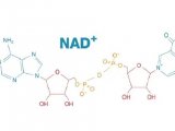
Figure 1. (Middle) UA, a bifunctional protein−DNA hybrid molecule, which binds to most types of IgG antibodies (left) and any DNAmodified reporter molecules (right) to generate a modular library of pre-labeled primary IgG antibodies for any applications of protein detection in place of secondary antibodies.
2.UA的合成与鉴定

(a) Scheme of forming the universal adapter (UA) using a self-catalyzing enzyme (SNAP). Thiol-modified DNA tag was first conjugated to maleimide-benzyl guanine (BG) that served as the substrate for the SNAP enzyme. This BG-modified DNA tag was then linked to EZZ protein through SNAP catalysis to create the universal adapter (UA), a bifunctional protein-DNA hybrid molecule. (b) Confirmation of UA on 4-20% SDS-PAGE gel electrophoresis. The same gel was first stained with GelRed dye (left) to visualize DNA products and then stained with Comassie Brilliant Blue (right) to visualize protein products. Only UA with both DNA and protein components is visualized in both cases. Lane 1. UA; Lane 2. thiol-modified DNA only; 3. EZZ-SNAP protein only.
3.UA的功能验证---既能结合IgG,又能结合DNA修饰的标记分子

(a) Scheme to confirm the binding of UA against DNA nanobarcodes and IgG primary antibodies. DNA linker of UA was hybridized with DNA nanobarcodes to form UA-DNA nanobarcodes. UA-DNA nanobarcodes bound to IgG antibodies to form IgG nanobarcodes. (b) Characterizing of the hybridization of UA and DNA nanobarcodes on native PAGE gel electrophoresis. The same gel with no staining (left) showed signals of distinct color ratios from DNA nanobarcodes, and with GelRed nucleic acid stain (right) showed signals from all DNA products. Lane 1. UA; Lane 2. R2G0-DNA nanobarcode; Lane 3. EZZ protein mixed with R2G0-DNA nanobarcode; Lanes 4-6. UA hybridized with R2G0-, R1G1-, and R0G2-DNA nanobarcodes, respectively. The shifted bands in lanes 4-6 showed that DNA nanobarcodes were linked to UA through DNA hybridization. (c) Characterizations of the binding of UA-DNA nanobarcodes to IgG antibodies using dot blot technique. Different IgG antibodies were spotted on PVDF membrane and incubated with the UA-DNA nanobarcodes. Dots 1-3. Rabbit polyclonal antibodies (anti-glucagon, anti-PDX-1, and anti-insulin) labelled with R2G0-, R1G1-, and R0G2-nanobarcodes, respectively. Fluorescence signals detected from dots 1-3 showed strong binding between the IgG antibodies and EZZ protein of UA. Dot 4. Goat polyclonal antibody (anti-PDX-1) labelled with R1G1-nanobarcode. When using a non-specific IgG that cannot bind with EZZ protein, there was no fluorescence signal detected from dot 4. Dot diameters were 250 Km for all images.
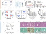
4.蛋白检测---利用IgG nanobarcodes,通过点杂交、微球和免疫染色的方式对相应蛋白进行检测

Figure 2. Using IgG nano-barcodes for protein detection. (a) Multiplexed protein detection with dot blot. Proteins were spotted on a PVDF membrane and then incubated with a detection solution, which contained a mixture of IgG nano-barcodes. The dots with fluorescence signals showed specific binding between protein targets and the corresponding IgG nano-barcodes. From left to right: GFP, RL, HSF, and sortase (negative control) proteins were labeled with R2G0-anti-GFP, R1G1-anti-RL, and R0G2-anti-HSF IgG antibodies, respectively. Dot diameters were 250 μm for all images. Background fluorescence from GFP alone was subtracted from the measurement with IgG nano-barcodes. (b) Multiplexed bead-based protein detection. PS beads were tagged with the first IgG antibody which captured the antigen and then sandwiched with IgG nano-barcodes. GFP, RL, and HSF protein targets were specifically detected on microbeads with R2G0-anti-GFP (red), R1G1-anti-RL IgG antibodies (orange), and R0G2-anti-HSF (green), respectively. (c) IgG nanobarcodes for immunostaining of insulin and glucagon proteins in a diabetic mouse pancreas tissue. Insulin was stained with R0G2-antiinsulin (green) antibody, and glucagon was stained with R2G0-antiglucagon (red) antibody. Intense yellow regions are the autofluorescence of contaminated red blood cells.
5.蛋白检测---利用IgG-UA-QDs和IgG-UA-HRP,通过点杂交和western blot方式对HSF蛋白进行检测

Figure 3. DNA-based protein detection system with different reporter molecules. (a) Protein detection using dot blot technique with quantum dots. Quantum dots were used to replace DNA nanobarcodes in our IgG nano-barcodes. HSF protein on PVDF membrane was recognized by its specific antibody and then labeled with UA-QDs. (b) Comparison of WB detection of HSF protein using DNA-modified HRP with IgG-UA (IgG-UA-HRP) (left) and traditional HRPmodified secondary antibody (right). Lanes 1−5, HSF protein detection using our IgG-UA-HRP at 0.25, 0.5, 0.75, 1, and 2 pmol; lanes 6−9, HSF protein detection using traditional WB method which utilized the HRP-modified secondary antibody at 0.25, 0.5, 0.75, and 1pmol.


 首页
首页


