实验概要
Cytokine ELISPOT has become a powerful routine tool for the analysis of disease- as well as vaccine-induced T-cell responses. The method is limited, however, in that only one cytokine at a time is assessed. Fluorospot is based on the principle of ELISPOT, but facilitates the analysis of single cells secreting several cytokines, e.g., polyfunctional T cells, suggested to be of protective importance in various infectious diseases. By detecting each cytokine with a specific fluorophore and analyzing differentially colored spots by fluorophore-specific filter systems, cells producing single or multiple cytokines are identified. Fluorospot maintains the simplicity and sensitivity of the ELISPOT while taking the analysis a step forward toward multiplex analysis.
实验原理
ELISPOT has, due to its high sensitivity and simplicity, proven valuable for assessing antigen-specific T-cell responses, both with regard to specificity and magnitude. One limitation of the regular ELISPOT method is, however, that only one cytokine at a time is measured. Still, in many settings, it is desirable to measure the production of multiple cytokines in a single well. For example, in studies of HIV, TB, and malaria infection or in the development of vaccines against these and other diseases, enumeration of antigen-specific T cells secreting, e.g., IFN-γ, may not yield a complete picture of the quality of the immune response. Recent studies in the field have highlighted the importance of polyfunctional T cells that secrete multiple cytokines. The ability of CD4 and CD8 T cells to respond to antigen with a combination of, e.g., IFN-γ, IL-2, and TNF-α, rather than only one of the cytokines, has been associated with enhanced protective immunity in viral, bacterial, as well as parasitic diseases (1–4).
An ELISPOT-based assay analyzing multiple cytokines would not only be useful for defining polyfunctional T cells, it may as well be used for simultaneous measurement of functionally distinct cell populations of various types, e.g., T-cell populations, predominantly secreting single key cytokines representing, e.g., Th1, Th2, Th17, or Tregs. The possibility to measure multiple cytokines simultaneously in the same well also has other advantages, such as a need for less sample cells, valuable, e.g., in studies of mucosa-derived cells or studies on newborns and children (5).
Over the years, efforts to broaden the ELISPOT technique to include staining with substrates of two colors have been made, first for B cells secreting different Ig isotypes (6) and later for analysis of cytokines (7). Although successfully used in several studies (8, 9), the dual ELISPOT technique can be technically challenging to perform, suffers from several inherent analytical difficulties, and is limited to the analysis of two cytokines.
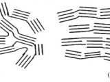
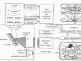
Building on the principle of ELISPOT, but using detection based on fluorescence instead of substrates, the fluorospot assay was developed (10). At present, two-color fluorospot has been described in several publications and reagents/kits for various cytokine combinations and species are commercially available. The most common protocol for fluorospot includes the use of biotinylated detection antibodies for one cytokine and an FITC-labeled detection antibody for the other cytokine (Fig. 1). As a second step in the detection, streptavidin conjugated to a red fluorophore (Cy3) and anti-FITC antibodies labeled with a green fluorophore, respectively, are used (10, 11). The resulting spots are subsequently analyzed using an automated reader equipped with filters for FITC and Cy3.

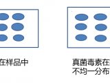
Fluorescent detection can be as sensitive as ELISPOT, or even more sensitive, and offers several advantages compared to dual ELISPOT. Most prominently, by using readers equipped with several narrow-band fluorophore filters, spots derived from cells secreting multiple cytokines are identified by the colocalization of single-colored spots in an overlay analysis of images from different filters (Fig. 2). Importantly, this enables the analysis of not only two, but also three and potentially even more cytokines simultaneously.

Experimental systems for triple-color fluorospot have been described (12), but the method may need further development before commercial reagents become available. The major limitation is the availability of additional amplification systems compatible with, e.g., FITC/anti-FITC and biotin/avidin and, in particular, automated readers designed for the analysis of three-colored spots. One amplification strategy that has been evaluated is to use a third detection antibody from a unique species that can be detected by fluorophore-labeled species-specific anti-Ig antibodies, reactive only with the third detection antibody (12). However, in a wider perspective, this may be a limiting factor since most highly functional antibody pairs are derived from a few species. Another strategy being employed for triple staining is the use of additional tag/anti-tag systems other than FITC/anti-FITC (Fig. 1).
 首页
首页