细胞骨架是细胞运动、极性建立、形态发生等重要细胞生物学过程的基础,也在组织胚胎发育,神经网络建立等重要生理学过程中发挥了重要的作用,是细胞生物学研究的中心领域之一。近年来,国内从事细胞骨架的研究队伍日益壮大,并取得了一系列的优秀科研成果。
为加强国内细胞骨架领域的交流与合作,由中国细胞生物学学会“细胞结构与细胞行为分会”主办,北京市细胞生物学会和山东省细胞生物学会协办,中国海洋大学承办的“第二届中国细胞骨架前沿学术会议”将于2021年7月28-31日在山东省青岛市举行。
本会议的宗旨是为国内细胞骨架及其相关领域的学者搭建学术和技术交流的平台,通过口头报告和墙报等多种形式展现国内该领域的最高科研水平,促进学术交流和合作,推动相关科研工作的进展。
牛津仪器ANDOR 将会在本次会议做技术报告并在会议现场设有展位,在会议期间ANDOR带来细胞骨架解决方案,届时欢迎各位莅临展位,与牛津仪器ANDOR工程师面对面交流和分享经验。
01
PART
会议详情
会议时间:2021年7月28日-31日
会议地点:山东·青岛 黄海饭店
主办单位:中国细胞生物学学会细胞结构与细胞行为分会
协办单位:北京市细胞生物学会和山东省细胞生物学会
承办单位:中国海洋大学
会议具体日程和详情,以主办方公布为准
02
PART
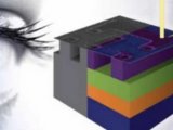
Dragonfly
多模式共聚焦成像系统

双转盘共聚焦/宽场/多色TIRF/dStorm
专利的BorealisTM完美照明系统
最高成像速度400fps-优于传统共聚焦10倍速度
光谱响应宽400nm-800nm
Micropoint专利激光活化照明系统
ClearView-GPUTM反卷积和Imairs原生数据格式
03
PART
Imaris
三维可视化图像分析系统
三维渲染与分割
细胞及其亚结构分析
丝状结构分析
活细胞轨迹追踪
多维共定位分析
04
PART
iXon Life EMCCD
单分子探测/超分辨成像黄金标准
单光子探测灵敏度
量子效率>95%
UltraVacTM专利真空密封
制冷温度低至-80℃
SRRF-Stream+超分辨技术-50nm
超高性价比
05
PART
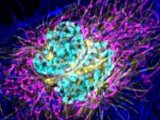
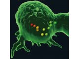
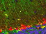
Dragonfly Gallery
以下图片均为Dragonfly成像
Adult zebrafish - Branchial cavity
Image Credit:Image acquired by Julien Rességuier at NorMIC (University of Oslo)
Adult zebrafish brain and vasculature
Image Credit:Image acquired by Julien Rességuier at NorMIC (University of Oslo)
Analysed Neuron

Image Credit:Image courtesy of Aubrianna Decker (Gaublomme Lab) and Daniel Virga (Polleux Lab)
Description: Neuron image taken using Andor sCMOS, Dragonfly, and analysed using Imaris 9.5. Image courtesy of Aubrianna Decker (Gaublomme Lab) and Daniel Virga (Polleux Lab).
Cell Biology(Confocal and SRFF)
Image Credit:Claudia Florindo (Andor) and Álvaro Tavares (CBMR, Universidade do Algarve)
Description: HeLa cells stained for MKLP1 (red), a-tubulin (microtubules in green), and DAPI (in blue). Image was acquired in a Dragonfly 505 with an iXON 888 camera using the SRRF-Stream+ mode.
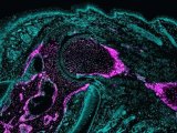
Organoid
Image Credit:Courtesy of Ronan Mellin, Dr. Luke Boulter, MRC Human Genetics Unit
Description: Staining- DNA (blue), LaminB1 (Green, nuclear envelope) and GM130 (Red, cis-golgi) Sample type- Mouse Colonic Epithelial Organoid.
PLA nanoparticles taken-up by endothelial cells
Image Credit:Image acquired by Julien Rességuier at NorMIC (University of Oslo)
Description: PLA nanoparticles taken-up by endothelial cells in Zebrafish.
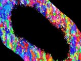
Zebrafish-Alessandra-Bromin
Image Credit:Courtesy of Alessandro Brombin (Patton Group)
Description: Zebrafish with red neural tube staining and MITFA-GFP cytoplasmic stain for melanocytes. 331 slices captured over 110um depth at 25x magnification with 25um pinhole. Zyla 4.2 There are very bright and very dim structures in the red channel. The 16-bit dynamic range of the camera mean all signals are detected and in range to that all detail is visible. A point scanning confocal finds this challenging to handle.
400 678 0609
Info.CHINA@oxinst.com


 首页
首页
