 返回 RESCue STED 超分辨活... 返回 RESCue STED 超分辨活...
 我要纠错
我要纠错
RESCue STED技术的基本原理
 RESCue
(REduction of State transition Cycles) STED is an improved,
photon-efficient STED imaging mode that significantly reduces the light
dose sent onto your sample without compromising the resolution – STED
imaging conditions designed for live-cell STED imaging!
RESCue
(REduction of State transition Cycles) STED is an improved,
photon-efficient STED imaging mode that significantly reduces the light
dose sent onto your sample without compromising the resolution – STED
imaging conditions designed for live-cell STED imaging!
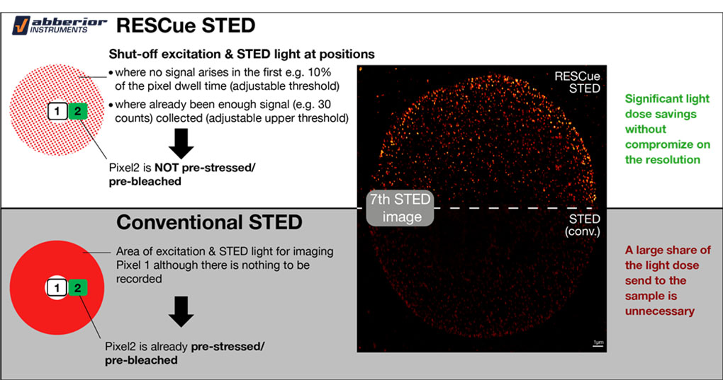
· RESCue avoids unnecessary excitation and de-excitation cycles and thus reduces photobleaching of any fluorescent marker
· The reduction in excitation and de-excitation cycles is particularly beneficial for volume imaging with 3D STED as well as for time lapse STED imaging
· RESCue STED applies excitation and STED light only at positions where fluorescent markers (i.e. the target structure) is present. It shuts off the lasers at positions where exposure would not make sense (i.e. where no markers are found)
· The resolution of the image is maintained although the light dose is reduced down to 4 % compared to gated cw STED imaging and down to 20 % compared to pulsed STED imaging
· RESCue STED is a highly photon-efficient way of superresolution imaging while conventional STED applies the same light dose to any pixel no matter if markers (i.e. a target) are present or not
· RESCue can be applied with similar benefits also to confocal imaging
RESCue STED 与其他技术的曝光剂量比较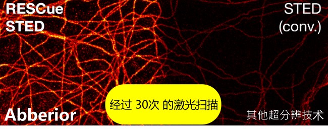 RESCue
STED 是 Abberior
STED 的附加模块, 可以不影响超分辨的光学解析下, 大副降低光辐射剂量, 仅是过去的gated-cw-STED 成像技术的 4% 光剂量而已. 在既有新一代的 Pulsed
STED 技术, 有了 RESCue STED 模块, 也仅是使用 20% 的光剂量而已.
RESCue
STED 是 Abberior
STED 的附加模块, 可以不影响超分辨的光学解析下, 大副降低光辐射剂量, 仅是过去的gated-cw-STED 成像技术的 4% 光剂量而已. 在既有新一代的 Pulsed
STED 技术, 有了 RESCue STED 模块, 也仅是使用 20% 的光剂量而已.
· RESCue STED 成像技术, zei适宜于活细胞超分辨成像及 3D-STED 的超分辨立体成像
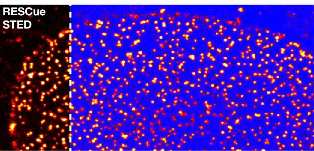 The
blue color indicates at which sample positions the excitation and the
STED laser were shut off in the RESCue STED imaging mode at the lower
threshold. Note that the blue positions correlate with the positions where no marker (i.e. target structure) is present.
The
blue color indicates at which sample positions the excitation and the
STED laser were shut off in the RESCue STED imaging mode at the lower
threshold. Note that the blue positions correlate with the positions where no marker (i.e. target structure) is present.
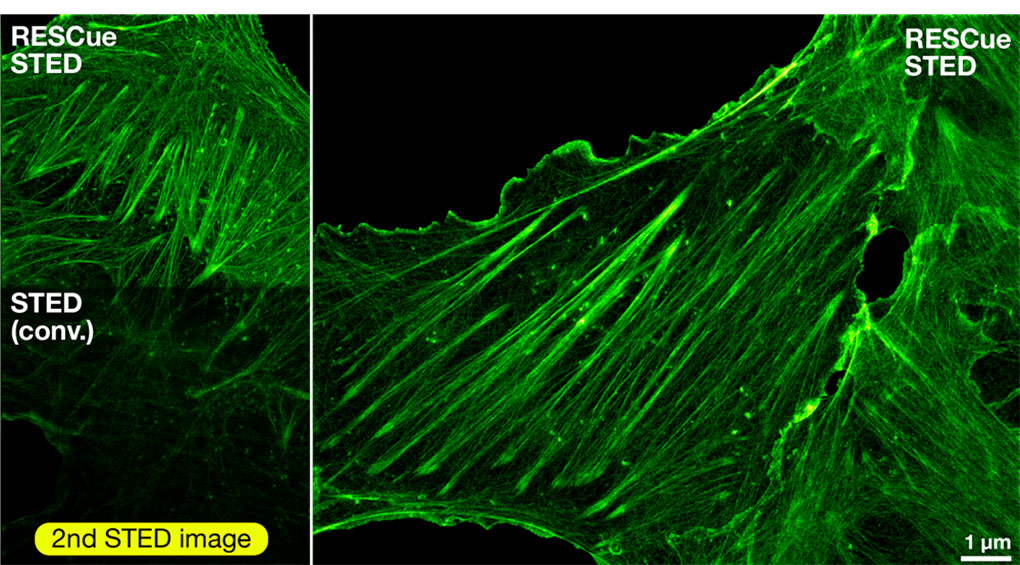
Shown
are Vero cells labelled with antibodies against nuclear pore complex
subunits in the central channel of the complex and secondary antibodies
coupled to Abberior STAR RED
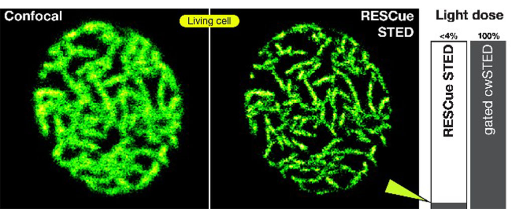 Shown
are Citrine-labelled eisosomes in living yeast cells (sample courtesy
of Prof. Dr. S. Jakobs, University Medical Center G?ttingen). Note that only ~4 % of the light dose was applied during image generation (compared to gated cw STED).
Shown
are Citrine-labelled eisosomes in living yeast cells (sample courtesy
of Prof. Dr. S. Jakobs, University Medical Center G?ttingen). Note that only ~4 % of the light dose was applied during image generation (compared to gated cw STED).
相较于 Leica 的 gated CW STED ( TCS SP8 STED ), Abberior RESCue STED 仅使用小于 4% 的光打到活细胞样本.
所以, 没有光漂白, 光毒害, 可以长时间做活细胞影像撷取
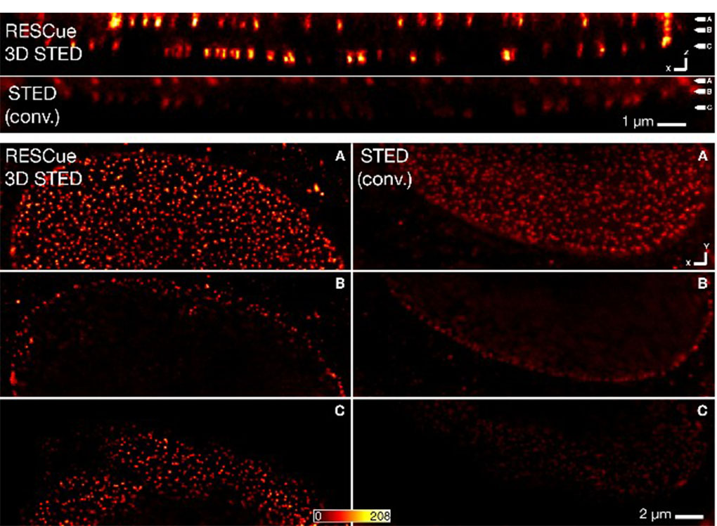 A
3D image stack of complete mammalian nucleus was recorded with RESCue
3D STED and conventional STED microscopy. The upper part of the figure
shows xz sections of a 3D STED image stack. The lower part shows three
planes (xy sections) of the 3D STED image stack. The position of the
individual xy planes is labeled by A,B,C in the top images.
A
3D image stack of complete mammalian nucleus was recorded with RESCue
3D STED and conventional STED microscopy. The upper part of the figure
shows xz sections of a 3D STED image stack. The lower part shows three
planes (xy sections) of the 3D STED image stack. The position of the
individual xy planes is labeled by A,B,C in the top images.
Shown are Vero cells labelled with antibodies against nuclear pore complex subunits in the central channel of the complex and secondary antibodies coupled to Abberior STAR RED

 参考成交价格: