作为一款操作简单的紧凑型台式拉曼成像系统,RA816生物分析仪将拉曼光谱的化学分析能力和先进的光学及光谱成像技术结合在一起,专为生物研究领域设计。RA816能够快速揭示生物样品的详细生化信息,包括组织活检、组织切片及生物流体等,具有高的灵敏度和特异性。
The Renishaw Biological Analyser is a compact benchtop Raman imaging system, designed exclusively for biological and clinical research. 紧凑的拉曼成像系统,专为生物和临床研究设计
Imaging and molecular medical diagnostic techniques can be sensitive and specific to information related to the initiation and progression of disease and pathology. However, these techniques require contrast agents (stains and labels) or molecular tags, which can be costly in time (processing and review expertise) and money.
Use the Renishaw Biological Analyser to identify and assess biochemical changes associated with disease formation and progression. There is little to no sample preparation, and no contrast agents or tags are needed.
The system provides a practical solution for analysing biological samples:
Easy to use hardware and software
No need for stains or labels
Minimal to no sample preparation
Obtain full range of biochemical information (no need for prior knowledge of specific molecular targets)
The system is designed specifically for biological and clinical sites:
Compact and transportable
Optimised microscopic technology:
Autofocusing (LiveTrack™ focus-tracking technology)
View samples at low and high magnification with minimum effort
Dedicated disease classification model building and testing
Dedicated tissue accessories and substrates
Designed for disease model transferability between instruments

Raman image of human osteosarcoma (bone cancer) cells. Raman image of human osteosarcoma (bone cancer) cells, showing the nuclei (green), nucleoli (red), membrane bound organelles (cyan) and the cell body (yellow – thick region, blue – membranous area). Renishaw thanks Dr Frederick Coffman, Rutgers New Jersey Medical School, USA, for providing the cell sample.

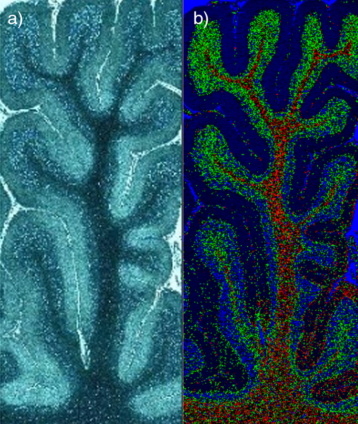
Imaging of human brain tissue Imaging of human brain tissue. Comparison of (a) white light and (b) Raman-false-coloured composite score image of cerebellum whole follicle showing arbor vitae/white matter (red), granule cell layer (green), molecular layer (dark blue) and meninges (pia, arachnoid and dura mater) (cyan)

Raman images of rat mandible (a) 2D and (b) 3D Raman images showing variation in surface topography of a rat mandible, as determined using Renishaw LiveTrack technology.