Live imaging provides an important complementation to the "snapshot" view obtained in fixed tissue by immunofluorescence. It allows following dynamic cellular processes as they unfold "in vivo", and often reveals a degree of complexity impossible to study in still images. Thanks to the increasing interest in this technique, significant technical improvements have been made in the recent years in microscopy leading to more sensitive and faster cameras, more efficient filter sets and a variety of different microscope systems to choose from. The temporal dimension of cellular processes in microscopy is still often neglected due to the difficulties in technical setup and the necessity to fabricate your own tools. Now the majority of the required equipment is commercially available from microscope manufacturers and related companies including environmental chambers, CO2-control and temperature control.
Often used in conjunction with time-lapse microscopy are fluorescent proteins, such as the Green Fluorescent Protein (GFP) or the Red Fluorescent Protein (RFP; Shaner et al., 2004; Shaner et al., 2005; Wiedenmann et al., 2004) and live dyes. Likewise this is a rapidly developing field of research, which has provided a variety of different "flavors" of fluorescent proteins for the scientific community, with properties specifically suited for different live-imaging applications (for a guide to choose the right one, see Shaner et al., 2005).
Back to top
Growth conditions, transformation and selection of stable cell lines are as described (Cherbas et al., 1994) using 100µg/ml hygromycin as a selection reagent. The following protocols can be used with most Drosophila tissue culture cells as long as they are semi-adherent.
I tested a variety of different Drosophila Schneider S2 cells for their growth behavior and found that most of them had some difficulties undergoing and completing cytokinesis, producing membrane bubbles and remaining attached for a long time. These phenotypes were least pronounced in a Schneider S2 cell line called L2-4, which I have used for most applications (obtained from the Botchan Lab, 171 Koshland Hall, UCB, Berkeley, CA, 94720, USA).
Fluorescent proteins (FP's) like eGFP (CLONTECH) represent a good choice for the green and mCherry for the red channel (Shaner et al., 2004). Depending on the properties of the protein of interest, the FP's can be used as an N-terminal or C-terminal fusions and in general are non-toxic to the cells.
FP's fused to histones (e.g. Histone H2B) are a good alternative for a chromosome counter stain. Hoechst 33342 and Draq5 are DNA specific live dyes, that don't stain significantly other nucleic acids. Unfortunately they are toxic to Drosophila tissue culture cells after prolonged exposure (> 2 hours) at concentrations that give a reasonable signal to noise ratio. Moreover the UV light to excite Hoechst 33342 dye is also damaging to the DNA in contrast to the lower wavelengths used for the FP's or Draq5.
Although live imaging can be performed on transiently transfected cells, a significant proportion of cells displayed tri-polar spindles in mitosis (~30% vs. 5% in untransformed or stable cells), thus it is advisable to use stably selected cell lines for studies in mitosis.
Cells are plated at 1x10⁶/ml in Schneider's Medium (GIBCO/Invitrogen), 10% FCS, Antibiotics (Sigma A5955; used at 33x instead of 100x) at 25°C and used two days later, when the cell culture should be exponentially growing with a density of ~3x10⁶/ml (generation time is ~23 hours).
This is a simple method for live imaging of Schneider S2 cells for short periods of time (< 6 hours), and can be used on both upright and inverted microscopes (see note 1, note 2 and comment 1):
22x22mm coverslips and special glass slides with a circular depression are cleaned with 70% Ethanol. Vacuum grease is applied to the rim of the circular depression of the slide. Due to the short time of imaging, sterility is less of an issue here;
10µl of cell suspension are added to the center of the coverslips;
The depression slide is put on top to "sandwich" the drop as described for the "hanging drop" method (see figure 1; comment 2; Heun et al., 2006; Shields and Sang, 1970);
Cells are allowed to settle for ~10 minutes before imaging.
For time-lapse imaging of cells over multiple days the following protocol can be used (only for inverted microscopes) (see note 3):
Culture dishes or slides with coverslip bottom are used. Some providers produce round culture dishes (e.g. Willco Wells B.V.®) or 8-well or single well culture slides (e.g. Nunc™ or ibidi®, see figure 2);
To follow cells for multiple days it is advisable to pre-treat the culture dishes or slides with Poly-L-Lysine to minimize cellular movement. In short the slides or dishes are incubated 30 minutes in 0.01% Poly-L-Lysine, drained and allowed to dry for 1 hour at 60°C or at RT overnight;
Particularly helpful for overnight experiments is the autofocus option. It is part of many microscope-controlling software (Metamorph®, Velocity®, softWoRx® etc.) and can be setup to refocus after longer time-intervals (e.g. once every hour). Also the cell tracking option can be useful if cells move a lot over time, although high cell density can confuse the program.
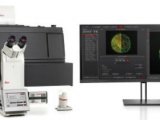
The protocol presented here is optimized for a Deltavision Spectris® and Deltavision RT® system using an inverted Olympus™ IX71 microscope. This microscope system is based on wide field microscopy. It uses an algorithm to calculate and remove the out-of-focus light in a process called deconvolution. The advantage of this and similar systems compared to Laser Confocal Microscopy is a reduced risk of photo damage to the cells and the possibility to follow a larger field of cells. In contrast these systems are limited for imaging objects with depths > 30µm, because of high amounts of scattered out-of-focus light. A comprehensive summary for different options of image acquisition and choice of microscope is given in protocol 15 on http://www.epigenome-noe.net/researchtools/protocols.php (Neumann et al.).
Time-lapse microscopy is done by taking z-stacks with step sizes from 0.2-1µm for each channel using an Olympus™ Planapo 60x oil-immersion objective for ~3 hours. Exposure times, number of z-sections and time-intervals between the image acquisitions depend on the signal to noise ratio of the fluorescence and the time resolution required to follow the cellular process of interest. These conditions have to be determined empirically for different applications. To follow a cell through mitosis I found it sufficient to take 7 z-stacks of 1µm step size for each channel once a minute. Under these conditions exposure time should not exceed more than 100 ms in the green (eGFP) and 50 ms in the red channel (mRFP) to reduce bleaching and photo damage (see note 4 and note 5) (an example of results that can be obtained upon observation times of about 3 hours is shown in figure 3).
Drosophila Schneider S2 cells are fairly sensitive to light. To observe cells for > 3 hours the number z-stacks or the exposure time have to be reduced or time-intervals between images should be increased empirically to minimize photo-toxicity. For longer periods of time-lapse analysis, I increased the time intervals to one z-stack every 5 minutes.
Images are deconvolved on a softWoRx® analysis workstation. If 3D information is not part of the analysis, deconvolved time-lapse movies are quick projected to facilitate the analysis in 2D and for display purposes. Bleaching of the FP's can be significant over time and often requires auto scaling to display the images with approximately similar brightness from beginning to end. Metamorph® allows this with very acceptable results, while other software's tested (softWoRx®) perform less convincingly.
For analysis in 3D, many software packages (Metamorph®, Bitplane Imaris®, Velocity®) allow the taking of a variety of different measurements such as the distance between two points of interest, volumes, distance traveled over time, etc. Whenever possible image analysis should be done as automated as possible to reduce the possibility of human error and bias towards expected results. A high image quality (= high signal-to-noise ratio) is a prerequisite for automated tracking, counting of objects and position information based on thresholding algorithms. However in reality lower image quality or high image complexity continues to present difficulties for commercially available image analysis software and needs to be done manually on a frame-to-frame basis.


 首页
首页


