Cyclopamine and jervine, teratogens derived from the skunk cabbage Veratrum californicum, are known to induce holoprosencephaly in chick embryos when introduced prior to gastrulation. Holoprosencephaly, a condition characterized by the reduction or apparent absence of left-right separation in certain tissues of the embryo, can be produced by a deficiency in the Sonic hedgehog signaling system. This disorder, also called cyclopia, results in offspring which are severely malformed and unable to survive due to serious brain defects.
Research performed by Michael K. Cooper and his associates showed that the presence of cyclopamine affects the signaling pathway of SHH in the early chick embryo. They claimed that cyclopamine acts as a class 2 transport inhibitor, a group of molecules which inhibit cholesterol esterification. These inhibitors seem to act by reducing the flow of cholesterol and its sterol precursors from the plasma membrane to the endoplasmic reticulum.
We planed on performing a study parallel to that performed by Cooper, only using Xenopus in place of chick embryos. Had we obtained similar results, then Xenopus and chick embryos may share common cues for signaling important information during development. This would illustrate that lower and higher vertebrates share common genetic programming.
By studying the growth of developing Xenopus embryos in cyclopamine solutions of varying molarity, obvious morphological mutations can demonstrate similarities between lower and higher vertebrates. If mutations are not found in the developing embryos, then SHH is yet another molecule whose function has changed throughout the course of evolution.
The cyclopamine used in this experiment was pre-dissolved in ethanol at a concentration of 1 mg per ml. With a molecular weight of 409.61 grams per mole, this is a 0.00244 molar solution. The calculations for determining the amount needed to reach a certain molarity of the Ringer's solution were fairly simple, though the ethanol had the potential to complicate the experiment.
To account for the toxicity of ethanol, this expermiment was conducted in two phases. In both phases the embryos (10 per sample for part 1, 15 per sample for part 2) were exposed to their designated concentrations of cyclopamine for 24 hours, at which time their Ringer's solution was replaced with fresh solution without the cyclopamine/ethanol mixture. The embryos were photographed every day as they developed.
In the first phase, the cyclopamine solution was used at varying concentrations (listed below in micromoles). This table showed that at any concentration greater than 100 micromoles, the embryos would not survive, so the second part of the experiment was changed to meet our new data.

In the second part of the experiment the potential effects of ethanol were examined by keeping a constant amount of ethanol in each sample. Using the data obtained from the first part of the experiment, we knew that at cyclopamine treatments above 100 micromoles there were no surviving embryos making these concentrations useless.
The following concentrations (in micromoles) were used, resulting in a certain number of mutations, deaths, and normal growths.
spacer | Control | 50 cyclopamine | 50 ethanol control | 100 cyclopamine | 100 ethanol control |
Normal | 15 | 6 w/ minor changes | 7 w/minor changes | 0 | 0 |
Mutated | 0 | 3 major | 3 major | 8 | 0 |
Dead | 0 | 6 | 5 | 7 | 15 |
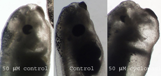
The addition of cyclopamine did not produce any cyclopic embryos, though mutations were seen in both the experimental and ethanol control groups. For example, this picture of 4 day old embryos shows changes in the eyes in both groups. While cyclopamine did not produce cyclopic embryos, it did produce more drastic changes.
 首页
首页


