
2021年9月29日 南方科技大学冷冻电镜中心 | ||
时间 | 主题 | 嘉宾 |
9:00-9:05 | 开场致辞 | 南科大和徕卡双方领导 |
9:05-9:10 | 徕卡示范实验室揭牌仪式 | 南科大和徕卡双方领导 |
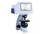
9:10-9:40 | 报告:徕卡冷冻电镜样品制备流程 | 徕卡显微系统 肖丽国 |
9:40-9:50 | 合影 | |
9:50-10:00 | 步行至南方科技大学冷冻电镜中心 | |
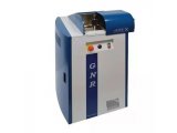
10:00-12:00 | 操作演示与上机实操 | 分三组 |
EM ICE | EM GP2 | CLEM/ ACE600/ VCT500 | |
10:00-10:40 | Group A | Group C | Group B |
10:40-11:20 | Group B | Group A | Group C |
11:20-12:00 | Group C | Group B | Group A |
12:00-13:30 | 午餐 | |
13:30-14:00 | 报告: Structure Determination of Excitation-Contraction Coupling Complex in Myocyte | 南方科技大学 刘铮 |
14:00-14:30 | 报告:眼见为实——低温透射电镜制样及相关技术介绍 | 同济大学 祝建 |
14:30-14:50 | 茶歇 | |
14:50-15:30 | 报告: Moving Beyond Particles in Cryo EM. High Pressure Freezing and Freeze Fracture, essential preparation tools for cryo SEM and cryo FIB. | 伦敦国王学院超结构成像中心主任 Roland Fleck |
15:30-16:10 | 报告:Leica Cryo CLEM Solutions – 有的放矢:获得目标结构高分辨数据 | 徕卡显微系统CLEM应用经理 Jan De Bock |
16:10-16:40 | 讨论与交流 | |
16:40-16:50 | 总结陈词 | 南科大冷冻电镜中心主任 王培毅 |

长按识别二维码
报名参与活动
特别说明:
本次活动全天直播
鉴于疫情管控因素,推荐广大校外学员线上参加研讨会
设置讨论与交流环节,欢迎广大学员积极提问
本次研讨会免费
活动主办方:


特邀报告人
刘铮
南方科技大学冷冻电镜中心 教授

1997年取得清华大学生物物理专业博士学位,2005年在美国Wadsworth Center任研究员,2015年兼同济大学医学院泛血管研究所副所长,心肺血管研究所副所长,博士生导师。2020年初入职南方科技大学冷冻电镜中心。
主要通过冷冻电子显微学、生物化学、细胞生物学等多种技术手段研究细胞器、膜蛋白等生物大分子的工作机制。先后在国内外重要学术期刊发表相关研究成果,其中SCI收录40余篇。

祝建
同济大学 教授
1982年毕业于宁夏大学农学院,留校任教
1992—1995年苏黎世瑞士联邦理工学院(ETH)细胞生物所电镜技术实验室,中瑞联合培养博士
1996—2000年上海铁道大学医学院
2000—2018年同济大学生命科学与技术学院,生物电镜技术实验室,教授,博导
ROLAND A. FLECK

上下滑动查看全部
Professor of Ultrastructural Imaging, Royal Society Industry Fellow and Director of the Centre for Ultrastructural Imaging at King''s College London, United Kingdom.
He is also a visiting Professor of the Faculty of Health and Medical Sciences, University of Copenhagen and Professor of the UNESCO Chair in Cryobiology, National Academy of Sciences of Ukraine, Institute for Problems of Cryobiology, Kharkiv, Ukraine. He joined King’s College London in 2013 from the National Institute for Biological Standards and Control (NIBSC), where he was head of Biological Imaging and Assay Development. At NIBSC he developed advanced imaging techniques for the control and standardisation of biological medicines and had research interests in developing differentiation protocols for myeloid leukemic and human embryonic stem cell lines as substrates for functional biological assays. He has extensive specialist knowledge of freeze fracture/freeze etch preparation of tissues and wider cryo-microscopic techniques.
As academic director of the Centre for Ultrastructural Imaging he collaborates widely with colleagues in neuroscience and parasitology and promotes advanced three dimensional studies of cells and tissues using both room temperature and cryo electron microscopy techniques. He has extensive experience and knowledge of low temperature biology and cryopreservation having researched how cells and tissues both avoid and are damaged by chilling and freezing events. His current research interests focus on developing tools and protocols for enhancing the preservation of tissues for characterisation by electron microscopy as a capacity to enhance wider scientific collaborations.
Areas of expertise: Advanced electron microscopy techniques, cryo electron microscopy preparation techniques and electron tomography, application of serial block face and focused ion beam for the life sciences.

Jan De Bock
上下滑动查看全部
Jan studied biology and did his PhD in the field of olfaction, characterizing olfactory neurons in their response to odorants.
Jan has worked as a microscopy expert in different roles since 2003. He joined Leica Microsystems in 2011 as a product specialist for confocal microscopy. In 2017, he became member of the newly formed Workflow and Application Team responsible for correlative workflows, in particular involving sample preparation and imaging under cryogenic conditions.
Abstract
Cryo Electron Microscopy workflows are a state-of-the-art tool to investigate proteins in the cellular context with subnanometer resolution. To succeed in this, light microscopes performing under cryogenic conditions are essential for an early quality check and to identify target structures for the subsequent EM analysis.
In this webinar we show Leica Microsystem´s cryo microscope solutions for the assessement of the sample quality, a safe sample transfer and super resolution imaging under cryogenic conditions as a basis for precise targeting.

冷冻电子断层图像。与核孔复合体相连的蛋白酶体(紫色)。由分子结构系Ben Engel博士提供。生物学,生物化学MPI,马丁斯利德,德国
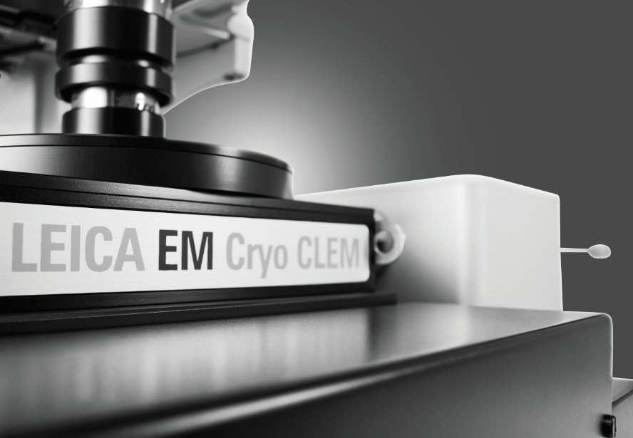
Leica EM Cryo CLEM
细胞内大分子原位结构研究
徕卡显微系统可为不同品牌SEM-FIB双束显微镜定制提供完全集成的低温电子断层成像工作流程,以满足研究需要。


 首页
首页
