Analysis of pigments often requires a slightly different use of a spectrophotometer. In the use of the instrument for determination of concentration (Beer-Lambert Law), the wavelength was pre-set and left at a single value throughout the use of the instrument. This value is often given by the procedure being employed, but can be determined by an analysis of the absorption of a solution as the wavelength is varied.
The easiest means of accomplishing this is to use either a dual beam spectrophotometer or a computer controlled instrument. In either event, the baseline must be continously re-read as the wavelength is altered.
To use a single beam spectrophotometer (such as the Spec 20), the machine is zeroed first, the wavelength is set, the blank is adjusted and then the sample is inserted and read. The wavelength is then adjusted up or down by some determined interval, the zero is checked, the blank re-inserted and adjusted, and the sample re-inserted and read. This procedure continues until all wavelengths to be scanned have been read.
In this procedure, the sample remains the same, but the wavelength is adjusted. Compounds have differing absorbtion coefficients for each wavelength. Thus, each time the wavelength is altered, the instrument must be recalibrated.
A dual beam spectrophotometer divides the light into two paths. One beam is used to pass through a blank, while the remaining beam passes through the sample. Thus, the machine can monitor the difference between the two as the wavelength is altered. These instruments usually come with a motor driven mechanism for altering the wavelength, or scanning the sample.
The newer version of this procedure is the use of an instrument which scans a blank, and places the digitalized information in its computer memory. It then rescans a sample and compares the information from the sample scan to the information obtained from the blank scan. Since the information is digitalized (as opposed to an analog meter reading), manipulation of the data is possible. These instruments usually have direct ports for connection to personal computers, and often have built in temperature controls as well. This latter option would allow measurement of hanges in absorbtion due to temperature changes (known as hyperchromicity). These in turn can be used to monitor viscosity changes, which is related to the degree of molecular polymerization with the sample. For instruments with this capability, the voltage meter scale has given way to a CRT display, complete with graphics and built in functions for statistical analysis.
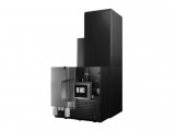
A temperature controlled UV spectrophotometer capable of reading several samples at pre-programmed time intervals is invaluable for enzyme kinetic analysis. An example of this type of instrument is the Beckman DU-70.
For routine use, substances to be monitored by spectrophotometry are often reacted with dyes to form a complex that is of another color, usually one easily read within the visible light range, and with precision by an instrument such as the Spec 20.
MATERIALS
Lyophilized bovine plasma gamma globulin or bovine serum albumin (BSA)
Coomasie Brilliant Blue 1
0.15 M NaCl
Spectrophotometer and tubes
Micropipettes
PROCEDURE (STANDARD ASSAY, 20-150 µ g protein; 200-1500 µ g/ml)
Prepare a series of protein standards using BSA diluted with 0.15 M NaCl to final concentrations of 0 (blank = NaCl only), 250, 500, 750 and 1500 µ g BSA/ml. Also prepare serial dilutions of the unknown sample to me measured.
Add 100 µ l of each of the above to a separate test tube (or spectrophotometer tube if using a Spec 20).
Add 5.0 ml of Coomasie Blue to each tube and mix by vortex, or inversion.
Adjust the spectrophotometer to a wavelength of 595 nm, and blank using the tube from step 3 which contains 0 BSA.
Wait 5 minutes and read each of the standards and each of the samples at 595 nm wavelength.
Plot the absorbance of the standards vs their concentration. Compute the extinction coefficient and calculate the concentrations of the unknown samples.
PROCEDURE (MICRO ASSAY, 1-10 µ g protein;
Prepare standard concentrations of BSA of 1, 5, 7.5 and 10 µ g/ml. Prepare a blank of NaCl only. Prepare a series of sample dilutions.
Add 100 µ l of each of the above to separate tubes (use microcentrifuge tubes) and add 1.0 ml of Coomasie Blue to each tube.
Turn on and adjust a spectrophotometer to a wavelength of 595 nm, and blank the spectrophotometer using 1.5 ml cuvettes.
Wait 2 minutes and read the absorbance of each standard and sample at 595 nm.
Plot the absorbance of the standards vs their concentration. Compute the extinction coefficient and calculate the concentrations of the unknown samples.
MATERIALS
0.15% (w/v) sodium deoxycholate
72% (w/v) trichloroacetic acid (TCA)
Copper tartrate/carbonate (CTC)
20% (v/v) Folin-Ciocalteu reagent
Bovine Serum Albumin (BSA)
Spectrophotometer and tubes
Micropipettes
PROCEDURE
Prepare standard dilutions of BSA of 25, 50, 75 and 100 µ g/ml. Prepare appropriate serial dilutions of the sample to be measured.
Place 1.0 ml of each of the above into separate tubes. Add 100 µ l of sodium deoxycholate to each tube.
Wait 10 minutes and add 100 µ l of TCA to each tube.
Centrifuge each tube for 15 minutes at 3,000 xg and discard the supernatant.
Add 1.0 ml of water to each tube to dissolve the pellet. Add 1.0 ml of water to a new tube to be used as a blank.
Add 1.0 ml of CTC to each tube (including the blank), vortex and allow to set for 10 minutes.
Add 500 µ l Folin-Ciocalteu to each tube, vortex and allow to set for 30 minutes.
Turn on and zero a spectrophotometer to a wavelength of 750 nm. Use the blank from Step 7 to adjust for 100% T.
Read each of the standards and samples at 750 nm.
Plot the absorbance of the standards vs their concentration. Compute the extinction coefficient and calculate the concentrations of the unknown samples.
NOTES
The Lowry method depends on the presence of tyrosine within the protein to be measured. The standard protein must contain approximately the same number of tyrosine residues as the sample, or the procedure will be inaccurate. If there are no tyrosine residues in the sample to be measured, the Lowry method of protein determination is useless, and use should be made of the Bradford assay. In general, the Bradford assay is the method of choice for protein determinations.
MATERIALS
Biuret Reagent
Bovine serum albumin (BSA)
Spectrophotometer and tubes
PROCEDURE
Prepare standard dilutions of BSA containing 1, 2.5, 5.0, 7.5 and 10 mg/ml protein. Prepare serial dilutions of the unknown samples.
Add 1.0 ml of each of the standards, each sample, and 1.0 ml of distilled water to separate tubes. Add 4.0 ml of Biuret reagent to each tube. Mix by vortex.
Incubate all of the tubes at 37 ° C for 20 minutes.
Turn on and adjust a spectrophotometer to read at a wavelength of 540 nm.
Cool the tubes from Step 3, blank the spectrophotometer and read all of the standards and samples at 540 nm.
Plot the absorbance of the standards vs their concentration. Compute the extinction coefficient and calculate the concentrations of the unknown samples.
NOTES
The Biuret reaction was one of the first for the determination of protein concentration. It remains as a rapid determination, but is not very accurate. It is useful during protein separation procedures since there are fewer salt interference reactions than with the Bradford or Lowry techniques. The color formed is stable for about 1-2 hours and consequently all spectrophotometer readings must be made as soon as possible after the incubation step.


 首页
首页


